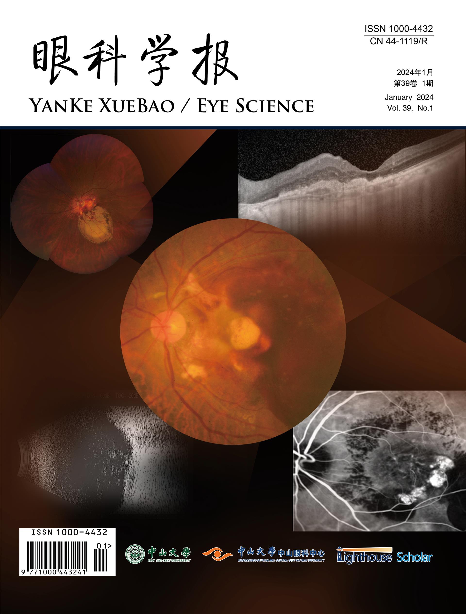
息肉状脉络膜血管病变(polypoidal choroidal vasculopathy,PCV)是亚洲人群中常见的致盲性眼病,发生大出血并发症后严重危害视力且预后差。PCV大出血包括视网膜下出血(subretinal hemorrhage,SRH)和玻璃体积血(vitreous hemorrhage,VH)。SRH的危险因素包括较长病程、簇型PCV、息肉状病灶不消退、合并视网膜色素上皮脱离;其治疗方式包括抗血管内皮生长因子药物、光动力疗法、激光、玻璃体腔注气、眼内注射组织纤溶酶原激活剂、玻璃体切割术或联合治疗等方式,其中,黄斑中心凹是否受累和出血时间是影响治疗方式选择的主要因素。发病年龄较大、白细胞计数较高、天门氨酸转移酶和丙氨酸转氨酶的比值较高、活化部分凝血活酶时间较长、曾行光动力疗法、有玻璃体腔注药治疗史、SRH面积大、出现视网膜色素上皮脱离的PCV患者发生VH的风险高。浓厚的VH通常需行玻璃体切割术,其手术时机和手术方式的选择是临床关注的焦点。鉴于目前PCV大出血的危险因素尚不完全明确、治疗方面也尚未形成共识,需要开展相关临床研究,提供更多依据。
Polypoidal choroidal vasculopathy (PCV) is a common blinding disease in Asian populations. Massive hemorrhage complications secondary to PCV includes subretinal hemorrhage (SRH) and vitreous hemorrhage (VH). The risk factors for SRH include a long duration, clustered PCV, non-regression of polyp lesions and presented with retinal pigment epithelial detachment. The treatments for SRH including anti-vascular endothelial growth factor drugs, photodynamic therapy, laser, vitreous pneumatic displacement, intravenously injected tissue plasminogen activator, vitrectomy and combination therapy. Whether macular fovea is involved and the time since bleeding onset are the main factors afecting the choice of treatment for SRH. Older age of onset, higher white blood cell count, higher aspartate amino transferase and alanine amino transferase ratio, longer activated partial thromboplastin time retinal pigment epithelium detachment, photodynamic therapy history, intravitreal injection history larger SRH area and presented with retinal pigment epithelial detachment were associated with higher risk of VH. PCV patients with massive VH should be treated with vitrectomy, while the timing and technique of operation should be paid atention to. At present, the risk factors of PCV massive bleeding are not completedly clear, and its treatment methods are diverse, which requires a large number of studies to prove its efectiveness and establish expert diagnosis and treatment consensus.
目的:探讨白内障人群角膜屈光力(corneal refractive power,CRP)的分布特点及与眼生物学参数的相关因素分析。方法:回顾性横断面研究福州眼科医院2019年3月至2022年7月就诊的40岁以上白内障人群共23035眼,使用OA-2000测量其眼轴(axial length,AL)、CRP、前房深度(anterior chamber depth,ACD)、晶状体厚度(lens thickness,LT)、角膜水平直径即白到白(white-to-white,WTW)、中央角膜厚度(central corneal thickness,CCT)。绘制各眼生物学参数及年龄Spearman相关性热力图,绘制CRP与AL、CRP与WTW散点拟合图。将CRP与上述参数及年龄进行Spearman相关性分析,分段数据的线性关系使用Pearson分析及线性回归分析。结果:白内障人群CRP为(44.36±1.52)D,在总体数据中CRP与AL为非线性相关;但在分段数据中存在线性相关:当AL≤25.06 mm,CRP与AL负线性相关(R2 =0.397,P<0.001);当AL>25.06 mm,CRP与AL正线性相关(R2 =0.045,P<0.001);无论AL长短,CRP与WTW、CCT均呈负相关。在总体数据中,CRP与WTW也存在非线性关系;但在分段数据中存在线性相关:当10.52 mm≤WTW≤12.46 mm,CRP与WTW负线性相关(R2 =0.149,P<0.001),并与AL、ACD、CCT呈负相关。结论:CRP与AL、WTW呈非线性相关,使用CRP优化计算人工晶状体(intraocular lens,IOL)屈光力时需适当考虑AL、WTW与CRP的相关性。
Objective: To investigate the distribution characteristics of corneal refractive power (CRP), and analyze the correlation between corneal refractive power and ocular biometric parameters in cataract patients. Methods: A retrospective cross-sectional study was conducted on 2,3035 eyes of cataract patients over 40 years old, who visited Fuzhou Eye Hospital during the period between March 2019 and July 2022. The subjects' examination results of axial length (AL), corneal refractive power (CRP), anterior chamber depth (ACD), lens thickness (LT), horizontal corneal diameter (WTW), central corneal thickness (CCT) were measured by OA-2000. Spearman correlation thermograms of bilological parameters and age for each eyes were worked out. The plot scatter fitting plots of CRP and AL, CRP and WTW were made. Spearman correlation analysis was made among CRP, above-mentioned parameters and age. Linear relationships of the segmented data were analyzed with Pearson and linear regression analysis. Results: In the cataract patients, CRP was (44.36 ± 1.52) D. There was a non-linear correlation between CRP and AL in the total data. However, there was a linear relationship in the segmented data. When AL ≤ 25.06 mm, CRP was negatively linearly correlated with AL (R2 =0.397, P<0.001). When AL>25.06 mm, CRP was weakly positively correlated with AL (R2 =0.045, P<0.001). Regardless of the length of AL, CRP was negatively correlated with WTW and CCT. There was also a nonlinear relationship between CRP and WTW in the total data. But there was a linear correlation in the segmented data.When 10.52 mm ≤ WTW ≤ 12.46 mm, the negative linear correlation was found between CRP and WTW (R2 =0.149, P<0.001), while there was negative correlation among CRP, AL, ACD, and CCT. Conclusion: There is a non-linear correlation among CRP, AL and WTW. To optimize the calculation of intraocular lens (IOL) refractive power with CRP, it is necessary to consider the correlation between AL, WTW, and CRP.
玻璃体切割术是目前临床上常见的眼科手术之一,其应用广泛,且具有良好的治疗效果,但术后仍会出现各种并发症,眼压升高便是其中常见的一种。玻璃体切割术后眼压升高的病因复杂多样,术前原发病的不同、术中处理方式的差异以及术后并发症均可引起眼压升高,根据不同的病因可以选用更合适的治疗方法。早期的眼压升高较易控制,主要采用药物及激光治疗,晚期眼压升高导致继发性青光眼则相对复杂,以手术治疗为主。该文主要对玻璃体切割术后高眼压的原因分析及治疗进展进行综述。
Pars plana vitrectomy is one of the common ophthalmic surgeries in clinic practice currently, which is widely used with good therapeutic effect. However, various complications may still occur after operation. Elevated intraocular pressure is one of common complications. The causes of postoperative ocular hypertension are complex and diverse. Elevated intraocular pressure could be caused by different preoperative primary diseases, intraoperative management methods,and postoperative complication. More appropriate treatment methods can be selected based on different causes. Early elevated intraocular pressure iseasier to control and is mainly treated with medicine and laser. Late elevated intraocular pressure leads to secondary glaucoma, which is relatively complex and mainly treated with surgery. This review mainly states causes and treatment progress of high intraocular pressure after vitrectomy.
目的:了解湿性老年性黄斑变性(age-related macular degeneration,AMD)患者自我感受负担(self-perceived burden,SPB)现状及其影响因素。方法:采用方便抽样法选取2021年1月至11月在中山大学中山眼科中心就诊的204例湿性AMD患者为研究对象,采用一般资料调查表、SPB量表、家庭支持自评量表、医学应对问卷对其进行测评。结果:患者SPB得分是(21.98±6.68)分,总体属于轻度SPB。湿性AMD患者的SPB水平与家庭支出(r=?0.326, P<0.001)和面对应对(r=?0.365, P<0.001)呈负相关,与回避(r=0.456, P<0.001)及屈服(r=0.310, P<0.001)应对方式呈正相关性。多重线性回归显示,独居、高龄、自费、双眼患病及采用回避应对的患者的SPB更高,而高文化水平、高家庭支持的患者SPB较轻。结论:湿性AMD患者有轻度SPB,但仍存在改善空间,医护工作者在工作中应重点关注高龄、文化程度低、家庭收入低、自费、独居、双眼患病及低视力的患者,及时进行心理疏导,减轻患者的SPB水平。
Objective: To understand the current status and influencing factors of self-preceived burden (SPB) in patients with wet age-related macular degeneration (AMD). Methods: 204 patiens with wet AMD who were treated in Zhongshan Ophthalmic Center, Sun Yat-sen University from January to November 2021 were enrolled as the study subjects with convenience sampling method. A general information questionnaire, SPB scale, family support self-assessment scale, and medical coping questionnaire were collected from the subjects for assessment. Results: The patient’s SPB score was 21.98±6.68, which is generally mild SPB. The SPB level of patients with wet AMD was negatively correlated with family support (r=-0.326, P<0.001) and coping (r=?0.365, P<0.001), and were positively correlated with avoidance (r= 0.456,P<0.001), and surrender (r=0.310, P<0.001) coping style. Multiple linear regression showed that the patients who lived alone, were elder and self-funded, had binoclur diseases and used avoidance coping, had higher SPB. While the patients with high education and family support had lower SPB. Conclusions: It is still needed to pay attention to the patients with AMD having mild SPB. Medical workers should focus on patients with elder age, low education level, low family income, self-funded, living alone, binocular disease and low vision in their work, and provide timely psychological counseling to reduce the SPB level of patients.
息肉状脉络膜血管病变(polypoidal choroidal vasculopathy,PCV)是亚洲人群中常见的致盲性眼病,发生大出血并发症后严重危害视力且预后差。PCV大出血包括视网膜下出血(subretinal hemorrhage,SRH)和玻璃体积血(vitreous hemorrhage,VH)。SRH的危险因素包括较长病程、簇型PCV、息肉状病灶不消退、合并视网膜色素上皮脱离;其治疗方式包括抗血管内皮生长因子药物、光动力疗法、激光、玻璃体腔注气、眼内注射组织纤溶酶原激活剂、玻璃体切割术或联合治疗等方式,其中,黄斑中心凹是否受累和出血时间是影响治疗方式选择的主要因素。发病年龄较大、白细胞计数较高、天门氨酸转移酶和丙氨酸转氨酶的比值较高、活化部分凝血活酶时间较长、曾行光动力疗法、有玻璃体腔注药治疗史、SRH面积大、出现视网膜色素上皮脱离的PCV患者发生VH的风险高。浓厚的VH通常需行玻璃体切割术,其手术时机和手术方式的选择是临床关注的焦点。鉴于目前PCV大出血的危险因素尚不完全明确、治疗方面也尚未达成共识,需要开展相关临床研究,提供更多依据。
息肉状脉络膜血管病变(polypoidal choroidal vasculopathy,PCV)是亚洲人群中常见的致盲性眼病,发生大出血并发症后严重危害视力且预后差。PCV大出血包括视网膜下出血(subretinal hemorrhage,SRH)和玻璃体积血(vitreous hemorrhage,VH)。SRH的危险因素包括较长病程、簇型PCV、息肉状病灶不消退、合并视网膜色素上皮脱离;其治疗方式包括抗血管内皮生长因子药物、光动力疗法、激光、玻璃体腔注气、眼内注射组织纤溶酶原激活剂、玻璃体切割术或联合治疗等方式,其中,黄斑中心凹是否受累和出血时间是影响治疗方式选择的主要因素。发病年龄较大、白细胞计数较高、天门氨酸转移酶和丙氨酸转氨酶的比值较高、活化部分凝血活酶时间较长、曾行光动力疗法、有玻璃体腔注药治疗史、SRH面积大、出现视网膜色素上皮脱离的PCV患者发生VH的风险高。浓厚的VH通常需行玻璃体切割术,其手术时机和手术方式的选择是临床关注的焦点。鉴于目前PCV大出血的危险因素尚不完全明确、治疗方面也尚未达成共识,需要开展相关临床研究,提供更多依据。
目的:了解干眼患者自我护理能力水平并分析其影响因素。方法:选取2022年2月—6月在中山大学中山眼科中心就诊的干眼患者为研究对象,采用一般资料调查表、自我护理能力量表、一般自我效能感量表对患者进行调查分析。结果:共调查293例干眼患者,其自我护理能力评分为(113.34±9.98)分,处于中等水平。相关性分析中干眼患者的自我护理能力总分与自我效能感得分呈正相关(r=0.421,P<0.001),多重线性回归分析显示,累计屏幕使用时间>10 h/d、合并全身疾病、低自我效能感评分是干眼患者自我护理能力的危险因素(P<0.05)。结论:干眼患者自我护理能力水平处于中水平,仍需加强。医护工作者在工作中应重点关注屏幕使用时间长、合并全身疾病及自我效能感低的患者,并制定相应的护理对策,以改善患者的自我护理能力水平。
Objective: To understand the self-care ability of patients with dry eye and analyze its infuencing factors. Methods: A total of 293 patients with dry eye were selected from Zhongshan Opthalmic Center, Sun Yat-sen University from February 2022 to June 2022, the general data Questionnaire the general self-efcacy Scale and the self-care ability Scale survey were collected. Results: A total of 293 patients with dry eye were surveyed, and the self-care ability score was 113.34±9.98, which was at the medium level. The total score of self-care ability, the scores of self-concept, self-care responsibility, health knowledge level and self-care skills of patients with dry eye were positively correlated with the scores of self-efcacy (r=0.421, allP<0.001).Multiple linear regression analysis showed that cumulative screen usage time>10 hours/day, comorbid systemic diseases, and low self-efficacy scores were risk factors for self-care ability in patients with dry eye (P<0.05). Conclusions: Te self-care ability of patients with dry eye disease is at a medium level, and still needs to be strengthened. Medical workers should focus on patients with prolonged screen usage, comorbid systemic diseases, and low self-efficacy in their work, and tailor relevant nursing strategies to improve their self-care abilities.
目的:了解干眼患者自我护理能力水平并分析其影响因素。方法:选取2022年2月—6月在中山大学中山眼科中心就诊的干眼患者为研究对象,采用一般资料调查表、自我护理能力量表、一般自我效能感量表对患者进行调查分析。结果:共调查293例干眼患者,其自我护理能力评分为(113.34±9.98)分,处于中等水平。相关性分析中干眼患者的自我护理能力总分与自我效能感得分呈正相关(r=0.421,P<0.001),多重线性回归分析显示,累计屏幕使用时间>10 h/d、合并全身疾病、低自我效能感评分是干眼患者自我护理能力的危险因素(P<0.05)。结论:干眼患者自我护理能力水平处于中水平,仍需加强。医护工作者在工作中应重点关注屏幕使用时间长、合并全身疾病及自我效能感低的患者,并制定相应的护理对策,以改善患者的自我护理能力水平。
Objective: To understand the self-care ability of patients with dry eye and analyze its infuencing factors. Methods: A total of 293 patients with dry eye were selected from Zhongshan University Zhongshan Ophthalmology Center from February 2022 to June 2022, the general data Questionnaire the general self-efcacy Scale and the self-care ability Scale survey were collected. Results: A total of 293 patients with dry eye were surveyed, and the self-care ability score was 113.34±9.98, which was at the medium level. The total score of self-care ability, the scores of self-concept, self-care responsibility, health knowledge level and self-care skills of patients with dry eye were positively correlated with the scores of self-efcacy (r=0.421, allP<0.001). Multiple linear regression analysis showed that cumulative screen usage time>10 hours/day, comorbid systemic diseases, and low self-efficacy scores were risk factors for self-care ability in patients with dry eye (P<0.05). Conclusions: Te self-care ability of patients with dry eye disease is at a medium level, and still needs to be strengthened. Medical workers should focus on patients with prolonged screen usage, comorbid systemic diseases, and low self-efficacy in their work, and tailor relevant nursing strategies to improve their self-care abilities.
孔源性视网膜脱离(rhegmatogenous retinal detachment,RRD)是一种严重威胁视力的眼部疾病,目前治疗手段以手术为主,手术方式主要有视网膜气体填充术(pneumatic retinopexy,PR)、巩膜扣带术(scleral buckling,SB)以及经睫状体扁平部玻璃体切割术(pars plana vitrectomy,PPV)。目前对于RRD手术术式的选择仍然存在争议,因此研究及制定RRD手术方式抉择的临床策略具有重要的临床意义。而临床上制定RRD患者手术方案往往与患者的年龄、视网膜脱离时间、裂孔的类型、位置、数量、大小等等临床因素有关,该文就影响孔源性视网膜脱离手术抉择的相关临床因素进行综述。
Rhegmatogenous retinal detachment (RRD) is a serious eye disease threatening vision. Surgery is main treatment currently, and surgery approaches include pneumatic retinopexy (PR), scleral buckling (SB), and pars plana vitrectomy(PPV). There is still controversy over the selection of RRD surgery approaches, so it is great significant to study and develop clinical strategies for RRD surgery approaches. The surgical plans for RRD patients are often related to clinical factors, such as the patient’s age, retinal detachment time, type, location, quantity, size, etc. This article reviews the related clinical factors affecting the surgical decision for rhegmatogenous retinal detachment.
目的:分析湖南地区汉族人群中2型糖尿病患者的人口学特征及生化指标,寻找糖尿病视网膜病变的高危因素。方法:釆用病例对照研究,统计湖南地区正常人群、2型糖尿病但无视网膜病变患者、2型糖尿病视网膜病变患者的人口学特征及生化指标的相关数据,进行成组t检验及logistic回归分析,探讨分析糖尿病视网膜病发生的易感因素。所有研究对象均为汉族。结果:对照组[非糖尿病(non-diabetes mellitus,NDM)组]和2型糖尿病未合并视网膜病变[(non-diabetic retinopathy,NDR)]组之间性别分布、年龄分布、BMI、舒张压、HbA1c、总胆固醇、高密度脂蛋白(high-density lipoprotein,HDL)、尿酸及总胆红素差异无统计学意义(均P>0.05)。NDM组中腹围、收缩压、空腹血糖、三酰甘油、肌酐和低密度脂蛋白(low-density lipoprotein,LDL)值均低于NDR组,差异有统计学意义(均P<0.05)。NDM组中BMI、腹围、收缩压、舒张压、空腹血糖、HbA1c、总胆固醇、三酰甘油、肌酐和LDL值均低于2型糖尿病合并视网膜病变组(diabetic retinopathy,DR)组,差异有统计学意义(均P<0.05)。NDR组收缩压、舒张压、HbA1c、总胆固醇、三酰甘油和肌酐值均低于DR组,差异有统计学意义(均P<0.05)。结论:收缩压超过150 mmHg,舒张压超过90mmHg,糖化血红蛋白超过9%,血清肌酐超过100 μmol/L,三酰甘油超过3 mmol/L均为糖尿病患者发生视网膜病变的高危易感因素。
Objective: To analyze the demographic characteristics and biochemical indexes of type 2 diabetic patients in Han population in Hunan, and to find the high-risk factors of diabetic retinopathy. Methods: The data of demographic characteristics and biochemical indexes of normal population, type 2 diabetic patients but without retinopathy and type 2 diabetic retinopathy in Hunan were analyzed. Group t test and logistic regression analysis were used to analyze the susceptibility factors of diabetic retinopathy. All the subjects were Han population. Results: There were no significant differences in gender distribution, age distribution, BMI, diastolic blood pressure, HbA1c,total cholesterol, high-density lipoprotein, uric acid and total bilirubin between the control group [non-diabetes mellitus (NDM) group] and the type 2 diabetic without retinopathy group [non-diabetic retinopathy (NDR)group] (all P>0.05). The abdominal circumference, systolic blood pressure, fasting blood glucose, triglyceride,creatinine and low-density lipoprotein in NDM group were all lower than those in NDR group, and the differences were statistically significant (all P<0.05). BMI, abdominal circumference, systolic blood pressure, diastolic blood pressure, fasting blood glucose, HbA1c, total cholesterol, triglyceride, creatinine and LDL in NDM group were all lower than those in type 2 diabetic retinopathy (DR) group, and the differences were statistically significant (all P<0.05). The comparison between the NDR group and the DR group showed that the values of systolic blood pressure,diastolic blood pressure, HbA1c, total cholesterol, triglyceride and creatinine in the NDR group were all lower than those in the DR group, and the differences were statistically significant (all P<0.05). Conclusion: SBP ≥150 mmHg,DBP ≥90 mmHg, HbA1c ≥9%, serum creatinine ≥100 μmol/L, triglyceride ≥3 mmol/L are the high-risk factors of diabetic retinopathy.
目的:了解原发性开角型青光眼(primary open angle glaucoma,POAG)患者视野缺损的进展情况,探讨其发生进展的相关危险因素。方法:回顾性分析2014年1月至2018年7月就诊于北京大学第三医院眼科并有至少4次视野检查的POAG患者。按照患者首次视野检查的平均偏差或平均缺损进行分期。将历次随访视野检查的平均偏差或平均缺损与时间进行线性回归分析,取其斜率(dB/年)。根据平均偏差或平均缺损的斜率将患者分为进展组与无进展组。分析患者视盘周围视网膜神经纤维层(retinal nerve fiber layer,RNFL)厚度损害位置、平均随诊间隔时间、基线视野分期等因素与青光眼视野缺损进展的关系。结果:共纳入128例患者(252只眼),其中129眼使用Octopus视野计检查随访,基线视野缺损值为(10.91±5.76) dB;123眼使用Humphrey视野计,基线视野偏差值为(–10.62±6.89) dB。视野缺损早、中、晚期的比例分别为26.19%、36.51%和37.30%。进展组31只眼(12.30%),无进展组221只眼(87.70%)。上下方RNFL都存在重度损害的患者,其视野缺损更易进展(P<0.001)。平均随诊间隔时间≤4个月的患眼,发生进展的比例高于平均随诊间隔时间>4个月的患眼(P=0.058)。基线视野分期、年龄、性别、总随访时间与视野缺损进展未见显著相关性。结论:青光眼患者的视功能损害出现恶化是普遍存在的。上下方RNFL均存在重度损害、随诊间隔时间短与视野缺损进展相关。视神经结构的改变与功能损害具有相关性,结构改变的方位对功能损害进展有提示功能。规律随诊对病情监测有重要意义,对于可能快速进展的患者,应缩短随诊间隔时间。
Objective: To investigate the progression of visual field defect in primary open angle glaucoma (POAG), and to explore the related risk factors for its progression. Methods: A retrospective analysis was performed on patients with POAG who had at least 4 visual field examinations in the Department of Ophthalmology, Peking University Third Hospital from January 2014 to July 2018. The visual field was staged according to the mean deviation or mean defect of the first visual field examination. Linear regression analyses of mean deviation or mean defect were performed against time, and corresponding regression slopes (in decibels per year) were calculated. Patients were divided into progressive and non-progressive groups according to the mean deviation slope or mean defect slope. The relationship between retinal nerve fiber layer (RNFL) thickness lesion location, mean follow-up interval, baseline visual field staging, and the progression of visual field defect in glaucoma were analyzed. Results: A total of 128 patients (252 eyes) were included. Among them, 129 eyes were followed up with an Octopus perimeter, and the average mean defect value of the baseline visual field was 10.91±5.76 dB; while the other 123 eyes were followed up with a Humphrey perimeter, and the average mean deviation value of the baseline visual field was –10.62±6.89 dB. The proportion of early, middle and late visual field defects was 26.19%, 36.51% and 37.30%. There were 31 eyes (12.30%) in the progressive group and 221 eyes (87.70%) in the non-progressive group. Patients with severe damage to both the upper and lower RNFLs had more visual field defects (P<0.001). Patients with an average follow-up interval ≤4 months had a higher rate of progression than those with an average follow-up interval >4 months (P=0.058).There were no significant differences in baseline visual field stage, age, gender, and total follow-up time between the progression and progression-free groups. Conclusion: Deterioration of visual function impairment is common in glaucoma patients. The progression of visual field defects is associated with severe impairments which are present both in the upper and lower RNFLs, and short follow-up intervals. Optic nerve structure changes are related to functional impairment, and the location of structural changes is suggestive of functional impairment progression.Regular follow-up visits are of great significance for disease monitoring. For patients who may progress rapidly, the follow-up interval should be shortened.